03 / Mechanisms
Neural Communication
The translation of biology into information processing. Neurons turn ion flow, membrane voltage, and neurotransmitter release into sensation, movement, memory, and thought.
A single neuron is not just an electrical wire. It is a living cell with specialized compartments for receiving, integrating, transmitting, and reshaping information across changing circuits.


A neuron is a specialized signaling cell
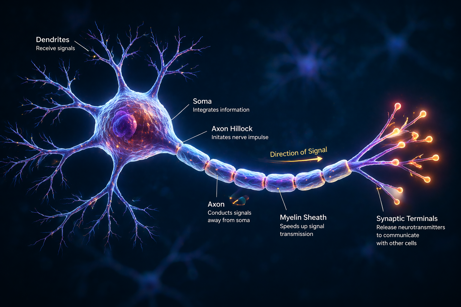
Neurons are polarized cells built for directional communication. Information usually arrives through dendrites, is integrated near the soma and axon hillock, and then travels outward along the axon toward synaptic terminals.
Neuron Anatomy At A Glance
Dendrites
Branch-like extensions that receive input from many other neurons. Their shape and receptor distribution influence what signals a cell can detect.
Cell Body
The soma maintains the neuron's metabolism and integrates incoming information. It houses the nucleus and much of the machinery needed to keep the cell alive.
Axon
A long projection that carries the action potential away from the cell body. Some axons travel only locally, while others connect distant brain regions.
Myelin
An insulating sheath wrapped by glial cells around many axons. Myelin speeds conduction and improves efficiency by allowing saltatory conduction between nodes.
Axon Terminal
The release site where electrical activity is translated into chemical signaling. Vesicles fuse here and spill neurotransmitter into the synaptic cleft.
From Input To Output
Neural communication unfolds as a sequence: receive input, integrate competing signals, fire when threshold is reached, then release transmitter onto the next cell.
Dendrites and the cell body receive thousands of excitatory and inhibitory inputs.
If the membrane reaches threshold near the axon hillock, the neuron generates an action potential.
The spike travels down the axon, often jumping between myelinated nodes to move faster.
At the terminal, calcium entry triggers neurotransmitter release onto the next cell.
The Action Potential Cycle
Because action potentials are all-or-none, neurons do not usually communicate stronger messages by making bigger spikes. They communicate by changing when they spike, how often they spike, and which cells spike together.
Excitation, Inhibition, and Signal Quality
Neural communication is not simply about turning neurons on. Functional circuits depend on an ongoing balance between excitation and inhibition. Excitatory input helps pass information forward, while inhibitory signaling shapes timing, suppresses noise, sharpens contrast, and prevents runaway activity.
This balance is one reason neural processing can be both flexible and stable. The brain must stay responsive to new input without becoming so reactive that information is lost in uncontrolled firing.

Why timing matters
Brains do not communicate only by sending more or fewer spikes. The timing of spikes across populations, the synchrony between regions, and the sequence in which signals arrive can all change what information a circuit extracts and how behavior unfolds.

The Synapse Is Where Signals Become Decisions
The synapse is the key conversion point between electrical and chemical signaling. One neuron releases neurotransmitter, but the next neuron does not simply copy the signal. It interprets that signal based on receptor type, membrane state, recent firing history, and the influence of nearby cells.
This is why two cells exposed to the same transmitter can respond differently. A synapse can amplify a message, dampen it, delay it, or make the circuit more likely to learn from it later.
Why glia matter too
Neural communication is not handled by neurons alone. Astrocytes help regulate ions and neurotransmitter levels, oligodendrocytes create myelin, and microglia contribute to maintenance and remodeling in changing neural environments.
Action Potentials
An action potential is the rapid electrical event that allows a neuron to send information over distance. When voltage-gated ion channels open, sodium ions rush in and depolarize the membrane. Potassium then flows out to repolarize it. Because this event is all-or-none, neurons encode many messages not by action potential size, but by timing and firing rate.
- The resting membrane potential is maintained by ion gradients and membrane permeability, especially to potassium.
- Threshold matters: only sufficiently strong summed input opens enough sodium channels to trigger a full spike.
- Refractory periods help set the maximum firing rate and ensure action potentials travel in one direction along the axon.
Synaptic Transmission
When an action potential reaches the presynaptic terminal, calcium enters the cell and triggers vesicles to release neurotransmitter into the synaptic cleft. That transmitter binds receptors on the postsynaptic membrane, changing the probability that the next neuron will fire. The result can be excitatory, inhibitory, slow and modulatory, or a combination of these depending on receptor type.
- Fast ionotropic receptors open ion channels directly and support rapid signaling.
- Metabotropic receptors act through intracellular cascades and often shape signaling over longer timescales.
- Transporters, enzymes, and glial cells help clear transmitter from the cleft and reset the synapse.
Neurotransmitters
Neurotransmitters are the chemical vocabulary of the nervous system. Glutamate is the main excitatory transmitter in much of the brain, while GABA is the main inhibitory one. Dopamine, serotonin, norepinephrine, and acetylcholine often act as modulators that shape motivation, attention, movement, mood, memory, and arousal depending on where and how they are released.
- The same transmitter can have different effects in different circuits because receptors and downstream signaling differ by cell type.
- Excitation and inhibition need to remain balanced; too much or too little of either can destabilize network function.
- Neuromodulators often change how circuits learn, prioritize information, or respond to reward and uncertainty.
Neuroplasticity & Learning
Neuroplasticity refers to experience-dependent changes in synaptic strength and circuit organization. One of the best-known examples is long-term potentiation, in which repeated coordinated activity strengthens synaptic transmission. Long-term depression weakens synapses under other conditions. Together, these mechanisms help neural circuits become more efficient at representing information and producing useful behavior.
- Plasticity is not limited to single synapses; it can also involve changes in dendrites, axons, and whole-network connectivity.
- Learning emerges when repeated activity changes what future activity becomes easier or harder for the circuit to produce.
- Plasticity also supports adaptation after injury, sensory loss, or intensive training.
Resources Used
These references support the educational summaries on this page and are included so readers can continue into primary or institutionally reviewed material.
Synaptic Transmission
Supports the step-by-step explanation of neurotransmitter release and postsynaptic signaling.
Neurotransmitter Synthesis
Used for descriptions of transmitter production, packaging, release, and removal.
The free-energy principle: a unified brain theory?
Provides theory background for prediction, learning, and adaptive brain signaling.